
Looking at the Bigger Picture - Combining hard X-ray and electron microscopy for correlative catalyst imaging
 Imaging techniques play a fundamental role in catalyst characterisation. They provide information on sample structure, composition and morphology with high spatial resolution and at different length scales.
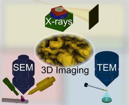
Transmission electron microscopy (TEM) is optimal for high resolution imaging down to atomic scale, but with strictly limited environmental conditions like gas and temperature. The sample may also be compromised by the need to prepare nanometre thin specimens, and the use of high energy electrons for imaging. On the other hand, hard X-ray microscopy (XRM) performed at synchrotron radiation sources can flexibly operate at elevated pressures or temperatures, and with micrometre to millimetre thick samples. Compared to electrons, hard X-rays are relatively non-invasive to the sample. However, spatial resolution is currently limited to some tens of nanometres. In reality, these techniques are highly complementary and the best of both methods can be combined using appropriate sample environments. XRM and TEM can provide not only imaging results, but also spectroscopic and diffraction data for a detailed understanding of the sample. One focal point is development of ‘in situ’ reactors, allowing catalyst imaging under relevant reaction conditions - the optimal way to observe catalysts at work and develop structure-activity relationships. Harnessing the power of these methods is a key research goal in the group.
Related publications
Y. Fam, T.L. Sheppard, A. Diaz, T. Scherer, M. Holler, W. Wang, D. Wang, P. Brenner, A. Wittstock, J.-D. Grunwaldt, ChemCatChem 2018, 10, 2858
S. Baier, C.D. Damsgaard, M. Klumpp, J. Reinhardt, T. Sheppard, Z. Balogh, T. Kasama, F. Benzi, J.B. Wagner, W. Schwieger, C.G. Schroer, J.-D. Grunwaldt, Microsc. Microanal. 2017, 23, 501.
S. Baier, A. Wittstock, C.D. Damsgaard, A. Diaz, J. Reinhardt, F. Benzi, J. Shi, T. Scherer, D. Wang, C. Kübel, C.G. Schroer, J.-D. Grunwaldt, RSC Adv. 2016, 6, 83031
Seeing is Believing - 3D catalyst characterization with hard X-ray tomography at the synchrotron
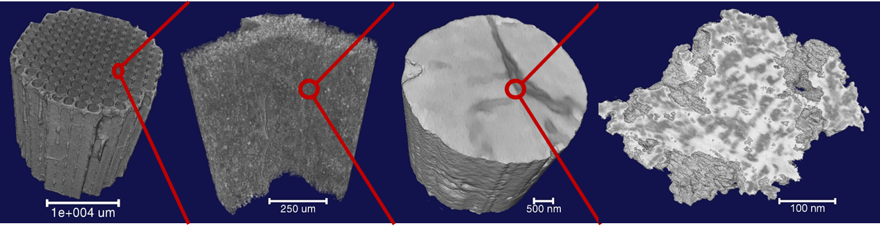
Heterogeneous catalysts are hierarchically-structured materials, which often show complex structural elements on multiple length scales. This includes metal nanoparticles or clusters, porosity (including micro-, meso- and macropores), and global structure or morphology, such as honeycombs, pellets or layers. Detailed characterisation of all these features and on all length scales is essential to develop understanding of catalyst form and function.
Hard X-ray micro- and nanotomography offers a versatile toolbox to meet this challenge. By taking advantage of the high flux, tuneable energy and sensitive detection capabilities of modern synchrotron radiation sources, it is possible to monitor the structure of catalysts in 3D. This can be combined with numerous contrast modes, including absorption (XAS), fluorescence (XRF), diffraction (XRD) or developing coherent methods such as ptychography. Furthermore, development of new sample environments even now permits 3D imaging in situ - under reaction conditions. These powerful methods are exploited in our group to look at everything from distribution and behaviour of metal nanoparticles, mapping of pore networks, and monitoring common catalyst deactivation processes such as sintering or coking. The many unexplored possibilities will surely lead to a bright future for X-ray tomography in catalysis research.
Related publications
P. Sprenger, T.L. Sheppard, J.-P. Suuronen, A. Gaur, F. Benzi, J.-D. Grunwaldt, Catalysts 2018, 8, 356
T.L. Sheppard, S.W.T. Price, F. Benzi, S. Baier, M. Klumpp, R. Dittmeyer, W. Schwieger, J.-D. Grunwaldt, J. Am. Chem. Soc. 2017, 139, 7855
G. Hofmann, A. Rochet, E. Ogel, M. Casapu, S. Ritter, M. Ogurreck, J.-D. Grunwaldt, RSC Adv. 2015, 5, 6893